1- introduction Gram Staining 2-Requirements Reagents 3-Method 4-Observations Contents. Bacteria that stain purple with the Gram staining procedure are termed Gram-positive.

The most widely used staining procedure in microbiology is the Gram stain discovered by the Danish scientist and physician Hans Christian Joachim Gram in 1884.
Gram staining lab report discussion. Gram Staining Lab Report Discussion. Gram Staining Lab Introduction Gram staining is a very important technique used in biology labs all over the world. It is a technique used to differentiate types of bacteria using certain physical and chemical characteristics Of their cell walls.
The Gram stain is a type of differential stain that allows a microbiologist to identify the differences between organisms andor differences within the same organism. Gram staining bacteria requires the use of aseptic technique to ensure the sterility of the experiment. The Gram stain is a type of differential stain that allows a microbiologist to identify the differences between organisms andor differences within the same organism.
Gram staining bacteria requires the use of aseptic technique to ensure the sterility of the experiment. Microbiology lab report. 1- introduction Gram Staining 2-Requirements Reagents 3-Method 4-Observations Contents.
4 The Gram staining The Gram staining method named after Hans Christian Gram. The Grams method are commonly classified as Gram-positive and Gram negative. The Gram stain is the most widely used staining procedure in bacteriology.
It is called a differential stain since it differentiates between Gram-positive and Gram-negative bacteria. Bacteria that stain purple with the Gram staining procedure are termed Gram-positive. Those that stain pink are said to be Gram-negative.
The terms positive and negative have nothing to do with electrical. INTRODUCTION Gram staining is the most essential and universally used staining technique in bacteriology laboratory. Gram-staining was firstly introduced by Cristian Gram in 1883This method is used to distinguish between gram positive and gram-negative bacteria which have consistent differences in their cell walls.
Gram-positive bacteria stain blue-purple and Gram-negative bacteria stain pink- red. The Gram stain is a differential stain used to identify two kinds of cells centered on the dissimilarity in their characteristics of staining reactions and cell wall anatomy under a microscope. Gram-positive stains purple and gram-negative stains.
In the first series of the analysis Gram staining was conducted in order to classify each of the three unknown bacterial strains are either Gram-positive or Gram-negative. The three unknown bacterial samples were also inoculated on microbiological differential culture media following the aseptic transfer technique. In this experiment 3 microorganisms were stained using crystal v iolet for 15 seconds and were viewed under a light microscope using oil immersion.
Bacillus subtilis image 1 was observed to be rod-shaped and some cells appear to have been forming chains. Staphylococcus aureus image 2 appeared very populated and dense. Micro Biology Lab Gram Staining Lab Report Purpose The purpose of this lab was to experiment with a staining technique called Gram Staining.
Gram Stains is used to identify and classify bacteria it is a differential stain that allows you to classify. We reviewed major errors in Gram stain reports from positive blood cultures to identify patterns and potential clinical impact. An introduction should include pertinent background information.
In this case the history of the Gram stain and its significance in microbiology are relevant 1 point. A discussion of the mechanism of Gram staining and how it differentiates bacteria on the basis of. Gram Stain Lab Report.
Culture I immediately made a streak plate and started the Gram Stain protocol1. In the first two slides I made and stained my results were ambiguous. Without putting the slide into the microscope I noticed one was pink and the other purple.
I hypothesize that in the cultures used in this lab for gram staining will contain many gram negative and gram positive bacteria. Material and Methods The following things are needed to run a gram staining experiment. Ammonium Oxalate-Crystal Violet stain 5.
Grams Iodine Solution 6. Gram staining method the most important procedure in Microbiology was developed by Danish physician Hans Christian Gram in 1884. Gram staining is still the cornerstone of bacterial identification and taxonomic division.
This differential staining procedure separates most bacteria into two groups on the basis of cell wall composition. A Gram stain test was done to the isolate using techniques covered in Lab 4. A Gram positive and Gram negative bacteria stains different colors purple for positive and pink for negative which can then be observed through the microscope to determine whether the particular isolate is a Gram positive or Gram negative bacteria.
Lab report nerixa portillo biol 2310 september 17 2017 introduction to simple stain and gram stain the study of microbiology is small organisms but the naked. Conclusion in conclusion gram staining method is. Conclusion In conclusion Gram staining method is widely use to differentiate bacteria between Gram positive and Gram negative.
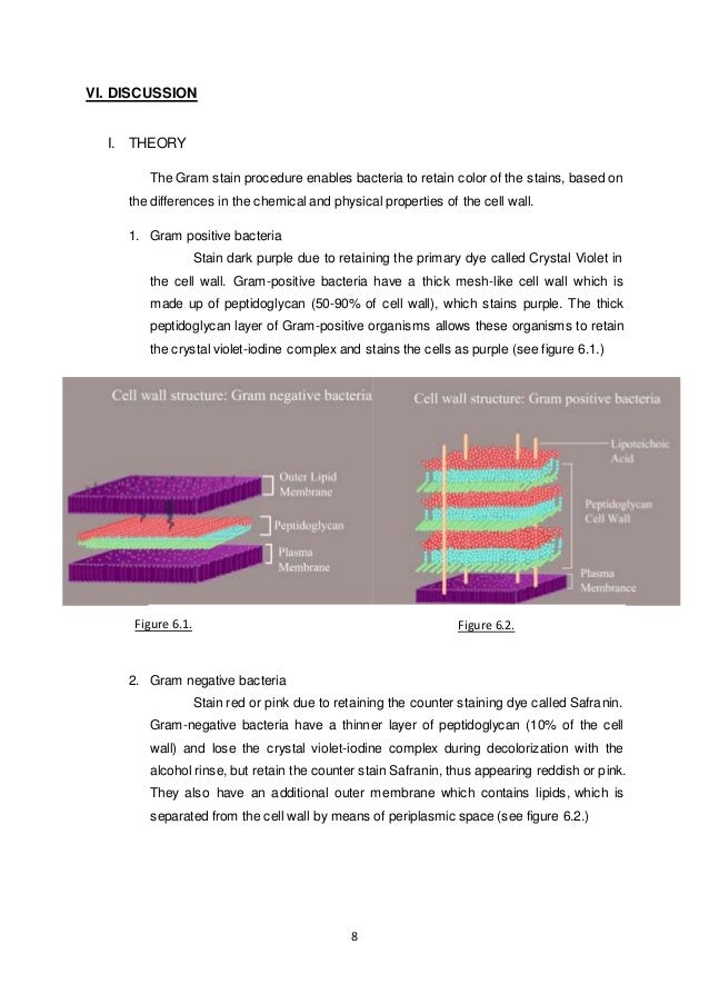
This methos will be great to bacteria that have cell wall but this method cannot cell acid fast bacteria which is bacteria that do not have cell wall. Gram positive bacteria with a thicker peptidoglycan layer retain crystal violet stain during the decolourisation process while Gram negative bacteria lose the crystal violet stain and are instead stained by the safranin in the final staining process6 From our observation from simple staining of Bacillus sp it can be seen that the bacteria is rod-shaped. The most widely used staining procedure in microbiology is the Gram stain discovered by the Danish scientist and physician Hans Christian Joachim Gram in 1884.
Gram staining is a differential staining technique that differentiates bacteria into two groups. The procedure is based on the ability of microorganisms to retain color of the stains used during the gram stain. The monochrome staining method is a direct stain that stains the bacteria using only one type of stain.
Gram staining method is useful for classification and identification of bacteria. The Gram stain is a differential stain which allows one to classify bacteria as Gram positive or. Gram Staining Formal Lab Report Miranda King Intro Microbiology Dr.
Hendrickson 13 September 2017 Purpose For the purpose of the gram staining experiment was to learn this specific type of staining technique as well as learn how distinguish the differences. 112 Abstract Escherichia coli Bacillus subtilis and Staphylococcus epidermidis were analyzed for this lab activity to determine their Gram Stain. After the multi-layered Gram Stain procedure each bacteria were classified as Gram-positive or Gram-negative depending on their cell walls staining color.
The results showed that E. Coli stained pink and classified as Gram-negative. In gram staining the bacteria cells are first stained with a primary stain crystal violet that enters into the cells and stain them purple-blue.
Then the cells are treated with a mordant grams iodine which enters into the cells and binds to the primary stain forming an insoluble dye-mordant complex crystal violet-iodine complex thus.